muscle fiber orientation
American Journal of Physiology - Heart and Circulatory Physiology. The angle of inclination of muscle fibers from coronal section was largest in the innermost and outermost zones and was progressively diminished toward the middle layer in all the hearts.

Fdi Muscle Fiber Tracks Derived From The Dwi Data In Three States At Download Scientific Diagram
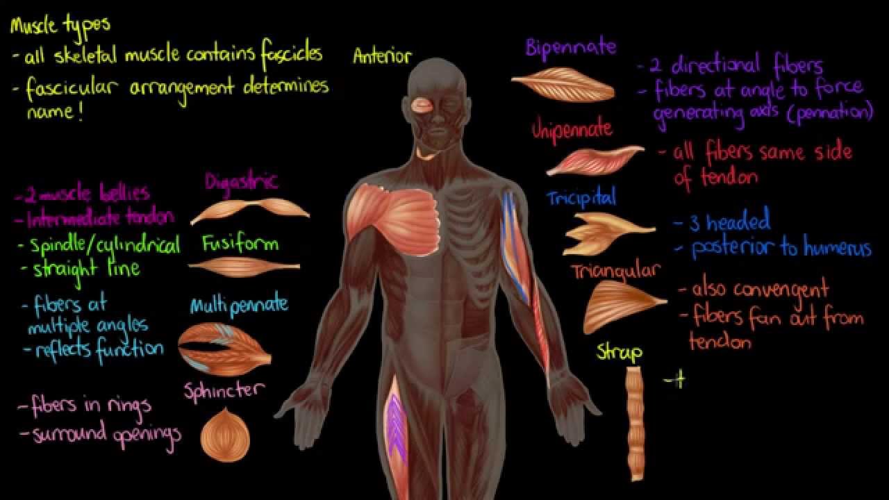
They are normally long muscles which cause large movements are not very strong but have good endurance.

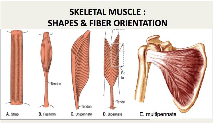
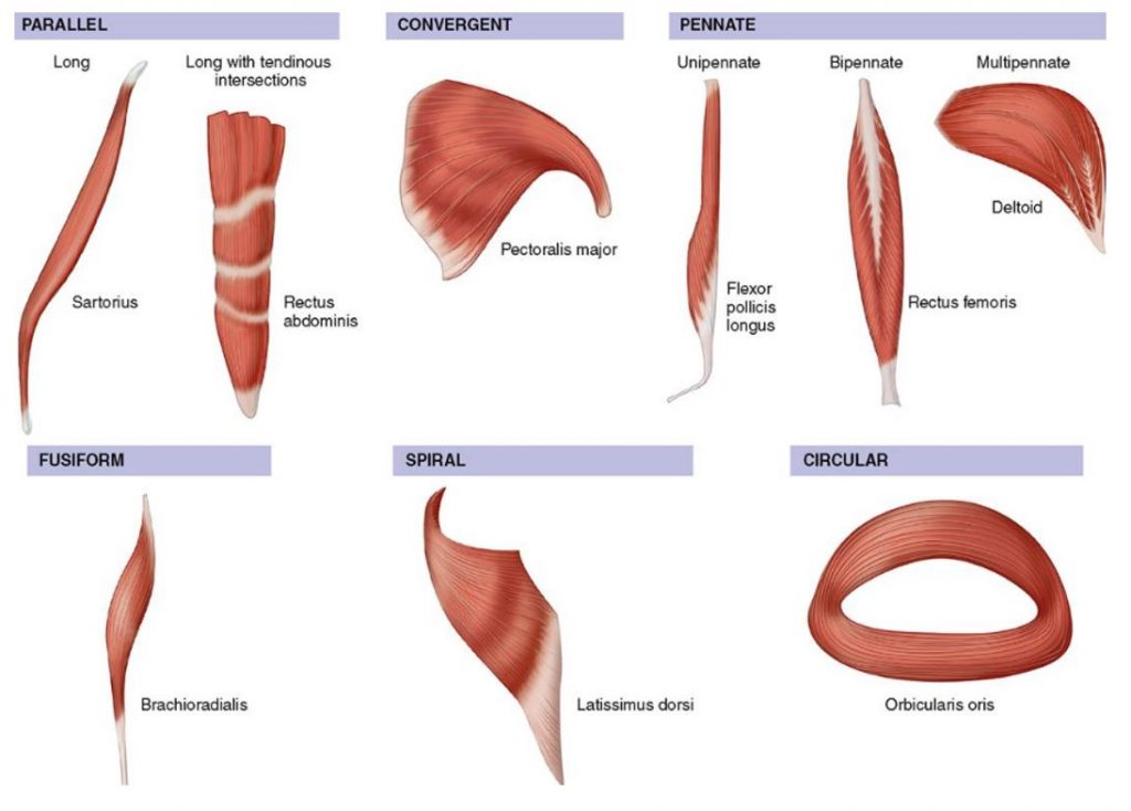
. Micromechanical modelling of skeletal muscles based on the finite element method. Examples include Sartorius and Sternocleidomastoid. Muscles with fibers arranged in a parallel orientation generally generate greater force.
280 - 20 gm. Group 1 five hearts. The endplate zone is assumed to be at about the midpoint of a muscle fiber.
Muscles with fibers arranged in series generally contracts more that is the distance between the proximal and distal tendons is reduced more than in muscles with fibers arranged in a parallel orientation. The bleaching correction step is based on a Macro by Sean McKinney. A muscle fiber orientation in which the component skeletal muscle cell is an equal distance apart for the entire length of the fibers everywhere.
From the 12 observations of the six animals which were conducted 15 and 21 weekspostoperatively fiber orientation II was much better than fiber orientation 1. The proximal and distal musculotendinous junctions in muscles of the upper and lower extremities were identified. Our research objectives are i to determine the fiber-oriented tensile response of skeletal muscle ii to simulate test responses using finite element analysis FEA for different fiber orientations and iii to compare the in vitro test results with the FEA results.
In fiberorientation II the musclefibersweregenerally in the circumferential direction of the heart perpendicular to the ventricular septum. The endplate zone is assumed to be at about the midpoint of a muscle fiber. This study was designed to locate the middle of the muscle fibers of commonly injected muscles thus identifying the endplate zone of these muscles.
The proximal and distal musculotendinous junctions in muscles of the upper and lower extremities were identified. This fiber orientation provides more range of motion but less power for contraction. PubMed Google Scholar Böl M Reese S.
Relating myocardial laminar architecture to shear strain and muscle fiber orientation. This project analyzes the orientation of muscle fibers in planarian flatworms. The endplate zone is assumed to be at about the midpoint of a muscle fiber.
The automatic methods proposed in recent years also involved voting procedures which were computationally expensive. This study reveals that the muscle orientation at 0 30 60 and 90 to the main magnetic field significantly affects the metabolite profile and quantification. This study was designed to locate the middle of the muscle fibers of commonly injected muscles thus identifying the endplate zone of these muscles.
Two groups of muscle fibers which are located within the deep perineal pouch and surround the urethra are called the urethral sphincter Oelrich 1980 The urethral sphincters are used to control the exit of urine in the urinary bladder through the urethra mainly the external urethral sphincter which is under voluntary control. Semitendinosus on the longitudinal cross section of the muscle. To elucidate the structural correlates of cardiac failure in myocardial tissue muscle fiber alignment and connective tissue volume fraction were measured at multiple sites in the left ventricular free wall and in the interventricular septum of 14 human hearts.
There are three different muscle fiber typesslow oxidative fast oxidativeglycoltic and fast glycolytic. The metabolite profile changes due to the muscle fiber orientation demonstrate that the positioning potentially causes inaccuracy in 1 H-MRS spectrum analysis. The dense muscle fiber microstructure gives rise to orientation dependent mr features with anisotropic overall motion of the creatine cr and phosphocreatine pcr molecules causing residual dipolar couplings first described for the total observed creatine tcr crpcr resonances 1 2 while orientation dependence was later also reported for.
Endurance training has minimal effect on the size of muscle however it does increase mitochondrial mass allowing for increased oxidative metabolism in skeletal muscle. The endplate zone is assumed to be at about the midpoint of a muscle fiber. The proximal and distal musculotendinous junctions in muscles of the upper and lower extremities were identified.
Some textbooks include Fusiform muscles in the parallel group. The scripts were developed for the Rink Lab at MPI-CBG in Dresden. A-g Within the same figure means lacking a common superscript were different P 005.
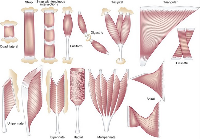
This study was designed to locate the middle of the muscle fibers of commonly injected muscles thus identifying the endplate zone of these muscles. Muscle fiber orientation in the left ventricular myocardial layer was histometrically estimated in normal concentric and eccentric hypertrophied hearts. Muscle fiber orientation STUDY PLAY parallel strap have fibres which as the name suggests run parallel to each other.
Scripts are written by Noreen Walker Scientific computing facility at MPI-CBG. Muscle fiber orientation varies between muscles and is dependent upon p ennation and muscle fiber length Christensen 1958 Saitou et. This study was designed to locate the middle of the muscle fibers of commonly injected muscles thus identifying the endplate zone of these muscles.
Muscle fiber orientation MFO is an important parameter related to musculoskeletal functions. Usage First run fiber_orientation_analysispy in Fiji. The proximal and distal musculotendinous junctions in muscles of the upper and lower extremities were identified.
Broadly there are two main approaches to extracting fiber orientationcurvature automatically from ultrasound. Muscle fiber orientation curvature and length is known to change with changes in force within the muscle Herbert et al. Muscle fiber orientation of m.
The traditional manual method for MFO estimation in sonograms was labor-intensive.

Pennate Muscle An Overview Sciencedirect Topics

Classification Of Muscles Based On The Directions Of Muscle Fibers The Download Scientific Diagram

A Unilateral Longitudinal Muscle Contraction Red Arrows Indicate Download Scientific Diagram

Fdi Muscle Fiber Tracks Derived From The Dwi Data In Three States At Download Scientific Diagram
Muscle The Primary Stabilizer And Mover Of The Skeletal System Clinical Gate

Myocardial Mechanics Structure And Function Of Myocardial Fibers Ecg Echo
Muscle Histology Flashcards Chegg Com

Organization Of Skeletal Muscles Course Hero
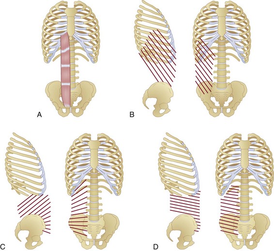
Anatomy And Mechanics Of The Abdominal Muscles Musculoskeletal Key

11 2 Explain The Organization Of Muscle Fascicles And Their Role In Generating Force Anatomy Physiology
Imaging Muscle Radiology Key

Marginal M Posterior P And Anterior A Fibers In The Soleus Download Scientific Diagram

Skeletal Muscle Shapes Fusiform Muscles Thick In Middle And Tapered At Ends Parallel Muscles Have Parallel Muscle Fibers Convergent Muscle Broad At Ppt Download

Pennate Muscle An Overview Sciencedirect Topics
Muscle Types Youtube
3 Orientation Of Cardiac Muscle Fibers Download Scientific Diagram

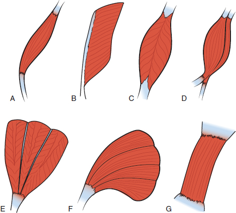
Five Possible Fiber Orientations In Intact Whole Muscle A Fusiform Download Scientific Diagram

Fascicle An Overview Sciencedirect Topics
How Muscles Work Part 2 Of 2 Shapelog


Comments
Post a Comment